The Zero Bone Loss Concept: A Path to Crestal Bone Stability

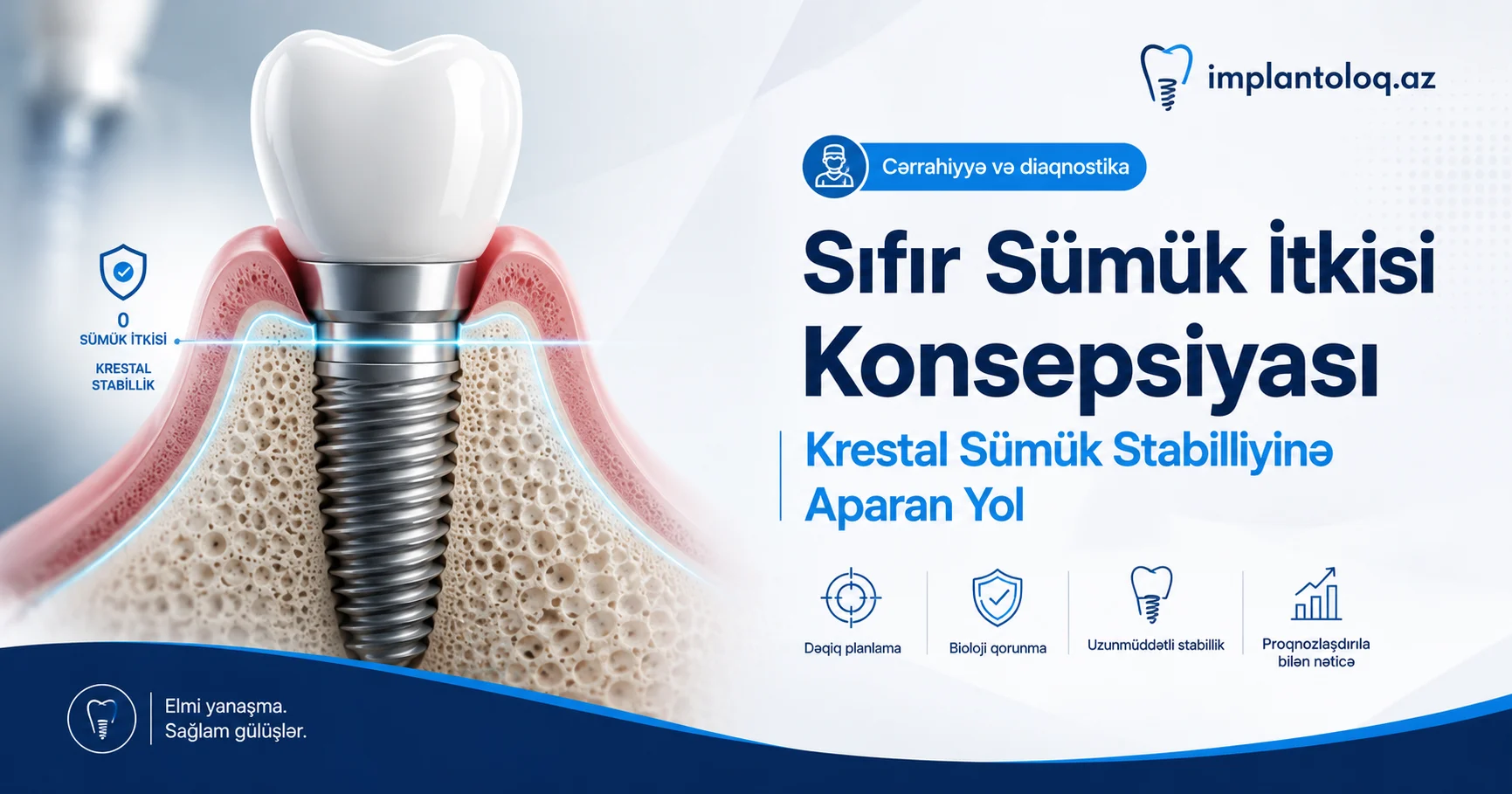
Keeping the crestal bone stable around an implant is one of the most important indicators of successful implant treatment. Even so, for decades crestal bone loss was considered an inseparable part of implantology, and for many clinicians it effectively became the norm.
One reason for this is the criteria published by Albrektsson and co-authors in 1986: according to them, bone loss of up to 1.5 mm during the first year after loading, and no more than 0.2 mm per year in the following years, is regarded as success. Because bone loss has so many possible causes, some clinicians believe it is impossible to control them all and give up on the idea of crestal bone stability altogether.
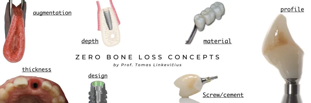
Professor Tomas Linkevičius and his research group consider this approach outdated and do not accept that bone loss is inevitable. For exactly this reason the Zero Bone Loss concept was developed: a set of clear clinical protocols designed to create and maintain crestal bone stability around implants.
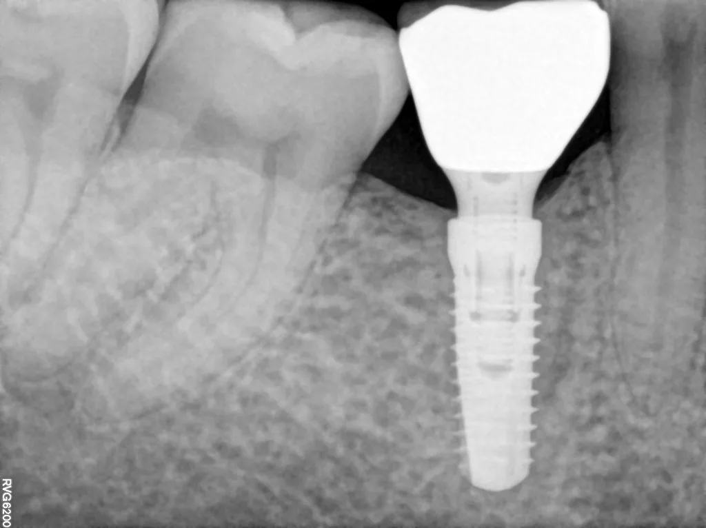
Stable marginal bone gives the patient a clinical advantage, and for the clinician it is also a source of peace of mind and professional satisfaction about long-term results. Every implantologist wants all of their cases to look like this years later.
That is why it is necessary to recognise the factors that can lead to loss of crestal bone stability and to use every available method to prevent bone resorption. But before listing the causes, one question needs to be answered: why is bone resorption so harmful for an implant? In the long term at least three serious problems arise.
Why is initial bone loss after 1 year accepted?
First, clinicians usually accept initial bone loss after the first year because, as a rule, this loss does not progress. But even if the bone level stays the same, a serious danger remains: if the area becomes infected again, the situation can turn into fairly severe peri-implantitis.
It should be kept in mind that a predictable treatment protocol for peri-implantitis has still not been fully established. For this reason the best treatment is prevention.
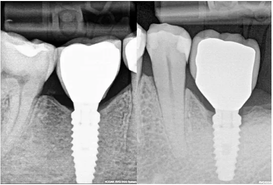
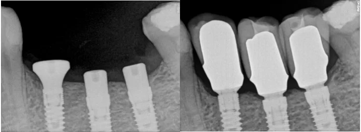
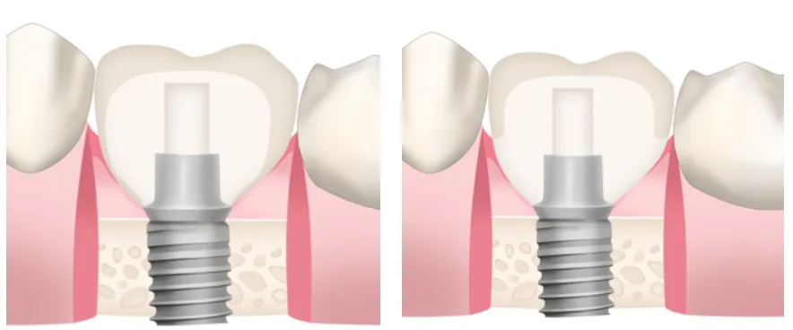
On the other hand, if we had the choice, each of us would choose the implant without bone loss (Figure 1A) rather than the one with bone loss (Figure 1B), however small the difference may seem. Sometimes clinicians play down the importance of this loss because they cannot explain why the bone was lost.
Bone loss and short implants: a dramatic connection
Second, crestal bone stability is especially decisive for short implants, which are being used more and more. Today an 8 mm implant is no longer considered short, and there is enough scientific data showing that 6 mm implants in the posterior regions of both jaws perform as well as long implants.
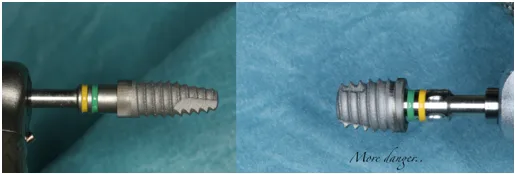
But imagine a 6 mm implant placed in the posterior mandible, where the vertical soft tissue is often thin. As the biological width forms, about 2 mm of bone resorption will occur. This means that only two thirds of the implant surface, the remaining 4 mm, stays osseointegrated. Taking into account the prosthetic construction on top and the implant/crown ratio, this is a real risk of losing the implant.
Some manufacturers even offer implants 4 mm long. For clinicians who use such products, soft tissue thickness is even more critical.
Height of the underlying bone
Third, the stability of the level of the peri-implant mucosa around the implant depends largely on the height of the underlying bone.
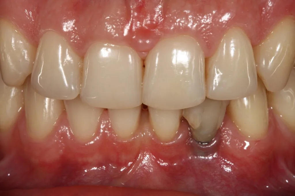
Migration of the marginal peri-implant mucosa as a result of early marginal bone loss seriously affects the aesthetics of the restoration, especially in the anterior region.
Soft tissue thickness: the key factor
So which factors matter for keeping the bone stable and preventing the complications listed above?
For about ten years the most effective solution to this problem was considered to be platform switching. It was regarded as so effective that almost every implant company made platform switching a "must-have" feature of production. As a result, the general conclusion took shape that implant design is more important than biology itself.
However, clinical studies by the Linkevičius group showed that soft tissue thickness is an important factor in maintaining crestal bone stability around implants.
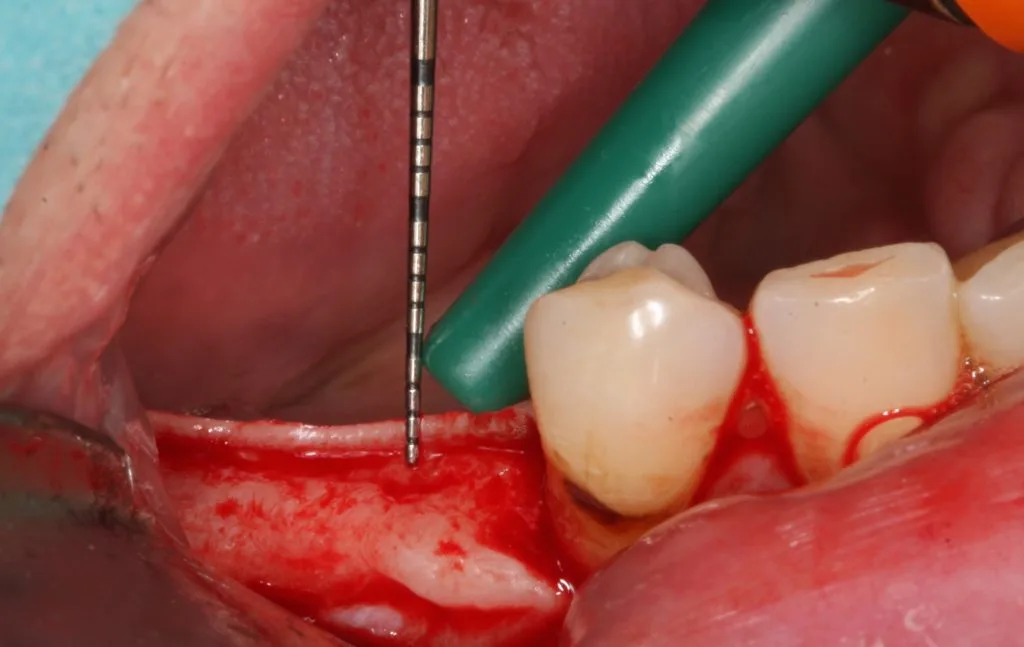
It was found that when the vertical soft tissue thickness is 2 mm or less, crestal bone resorption of about 1.5 mm occurs during the formation of the biological seal between the soft tissue and the implant/abutment/restoration surfaces.
Later studies showed that the minimum soft tissue height needed to prevent bone loss is 3 mm, and the optimal value is 4 mm, because 4 mm also allows a better prosthetic emergence profile of the restoration to be formed.
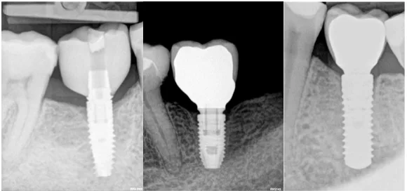
In addition, it has been clearly shown that if the vertical soft tissue is thin at the time of implant placement, even implants with a platform switching modification cannot preserve the bone.
This brings back the debate "which matters more: biology or implant design?" To take the question further, it is necessary to understand that vertical soft tissue thickness is the precondition for the biological width around the implant.
The biological width around an implant starts to form from the moment the healing abutment is connected and is fully completed after about 8 weeks. This biological seal is the only and most important barrier protecting the osseointegrated implant from the contaminated oral environment.
This means there is a direct link between the mucosa of the edentulous ridge before the implant and the peri-implant soft tissues. It appears that the soft tissue thickness needed to protect the bone around an implant is about 4 mm, which is more than the biological width around natural teeth.
There are only two ways for the biological width to form around an implant: with crestal bone loss or without bone resorption. Which one would you choose for your patient? Or, which one would you choose for your own mother? This is the question every clinician should answer honestly.
What is the strategy when thin tissue is found?
If thin vertical tissue is diagnosed at the time of implant placement, what should be done? Specific protocols have been developed in answer to this question, because otherwise crestal bone resorption is inevitable. There are several options: some have already been studied clinically, while others are based on clinical experience.
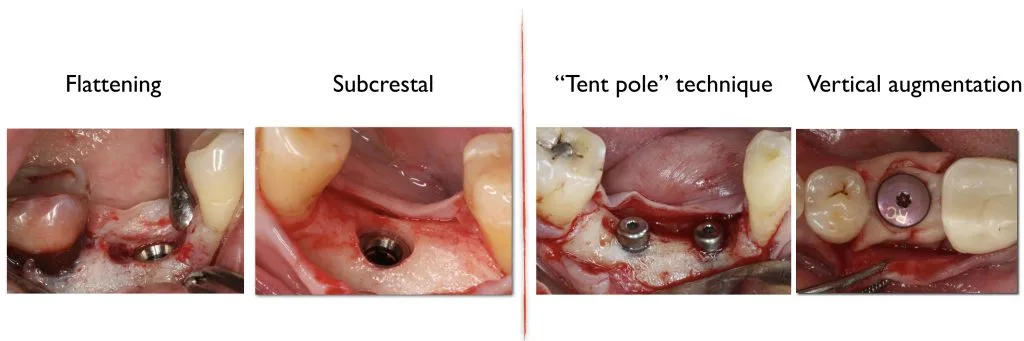
1. Subcrestal implant placement
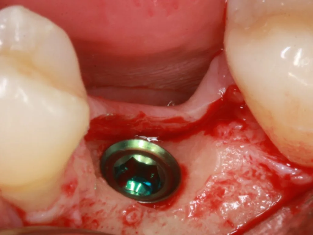
The first idea that comes to mind is to place the implant deeper, subcrestally. First of all, there must be enough distance to the alveolar nerve for the implant to sit safely in a subcrestal position; it is recommended that the implant sits at least 1 mm above the nerve.
Subcrestal placement leads not to bone loss but to controlled bone remodelling. Interestingly, not every implant can be placed subcrestally. In addition, when the implant is in a subcrestal position, the stability of the implant–abutment connection becomes especially important.
2. Bone recontouring
Another option is recontouring the bone during preparation of the main implant site, especially when the alveolar ridge is narrow.
Careful reduction and smoothing of a narrow alveolar ridge not only gives a flat bone surface and a wide enough area for placing the implant, but also increases the thickness of the soft tissue.
The concept of "removing bone to preserve bone" may seem unacceptable to some clinicians; also, a strong body of clinical evidence showing that this procedure increases soft tissue thickness and reduces crestal remodelling has not yet been established.
3. Vertical soft tissue augmentation
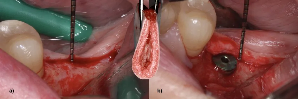
The third option is restoring soft tissue thickness in the vertical direction, and logically it is considered the most suitable approach: the vertical tissue deficiency is compensated directly.
This idea is not new. As early as 2009, the paper "The influence of soft tissue thickness on crestal bone changes around implants", published in the JOMI journal, recommended "considering thickening of thin mucosa before implant placement".
The goal is to increase the thickness of the soft tissue after healing by placing autogenous, allogeneic or xenogeneic material over the implant.
A connective tissue graft is considered the gold standard of soft tissue augmentation around implants. But this method has serious drawbacks: additional trauma at the donor site and the technical difficulty of harvesting the graft.
For this reason, soft tissue substitutes can be a real alternative to autogenous grafts in vertical reconstruction. A number of studies show that the vertical tissue deficiency that must be compensated for the biological width to form without bone loss can be made up at the time of implant placement using acellular dermal matrix material.
Prosthetic factors
The prosthetic component of the zero bone loss concept includes screw-retained restorations, titanium bases with a high gingival height, and polished zirconia in the subgingival area for the development of the epithelial attachment.
As a material, zirconia is considered the best choice for the peri-implant soft tissues. But it is important that it is made in a particular way, sufficiently polished.
It is also important not to make "zirconia without zirconia" restorations: when veneering ceramic completely covers the zirconia, direct contact between the soft tissue and the zirconia is not possible and the biocompatibility effect is lost.
Conclusion
The most important message is this: there is no single "most important" factor that ensures crestal bone stability. The result is determined by the combination and interaction of factors.
Once this multifactorial reality is accepted, the clinician can change the way of thinking and set out on the path to zero bone loss: correct diagnostics, assessment of soft tissue thickness, augmentation when needed and a precise prosthetic protocol.
If you are planning implant treatment in Baku, assessing the bone condition, soft tissue thickness and implant position with 3D tomography before surgery is a key condition for a long-term result. To book an appointment, you can contact us.
Source: Tomas Linkevičius, «Zero bone loss concepts. A path to crestal bone stability» — Periospot. This article is an adapted English translation.
This is general information. Your suitability, treatment time and cost are assessed individually during an examination.